Purine Bases, Pyrimidine Bases, Nucleosides and Nucleotides
Purine bases, pyrimidine bases and nucleosides were analyzed using RSpak DE-613 (a column for reversed phase chromatography).
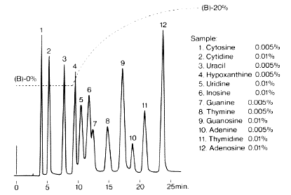
Sample :
1. Cytosine
2. Cytidine
3. Uracil
4. Hypoxanthine
5. Uridine
6. Inosine
7. Guanine
8. Tymine
9. Guanosine
10. Adenine
11. Thymidine
12. Adenosine
Column       : Shodex RSpak DE-613 (6.0mmID*150mm)
Eluent       : (A); 0.05M KH2PO4 aq.
               (B); 50% CH3CN
Flow rate    : 1.0mL/min
Detector     : UV(254nm)
Column temp. : 22deg-C
Six components were separated using RSpak NN-814 with pH3.0 phosphate buffer as the eluent. Since cation exchange mode is contributing to the separation, the elution order is different from that of the separation with RSpak DE-613.
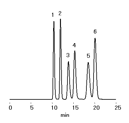
Sample : 20micro-L
1. Uridine 50micro-g/mL
2. Uracil 50micro-g/mL
3. Guanosine 100micro-g/mL
4. Cytidine 100micro-g/mL
5. Adenosine 50micro-g/mL
6. Cytosine 50micro-g/mL
Column       : Shodex RSpak NN-814 (8.0mmID*250mm)
Eluent       : 100mM NaH2PO4 aq./100mM H3PO4 aq.=500/66
Flow rate    : 1.0mL/min
Detector     : UV(260nm)
Column temp. : 25deg-C
Asahipak GS-320 HQ (a multimode column) is suitable not only for the separation of purine bases, pyrimidine bases, nucleosides and nucleotides but also for the separation of samples of their mixtures. GS-320 HQ permits one-time separation by isocratic elution without using gradient elution.
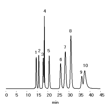
Sample : 20micro-L
1. ATP 100micro-g/mL
2. ADP 100micro-g/mL
3. IMP 100micro-g/mL
4. AMP 50micro-g/mL
5. GMP 100micro-g/mL
6. Inosine 100micro-g/mL
7. Adenine 100micro-g/mL
8. Hypoxanthine 100micro-g/mL
9. Guanine 100micro-g/mL
10. Adenosine 100micro-g/mL
Column       : Shodex Asahipak GS-320 HQ (7.5mmID*300mm)
Eluent       : 205mM NaH2PO4 aq./205mM H3PO4 aq.=300/7
Flow rate    : 0.6mL/min
Detector     : UV(260nm)
Column temp. : 30deg-C
Purine bases and pyrimidine bases were analyzed using Asahipak GS-320 HQ (a multimode column).
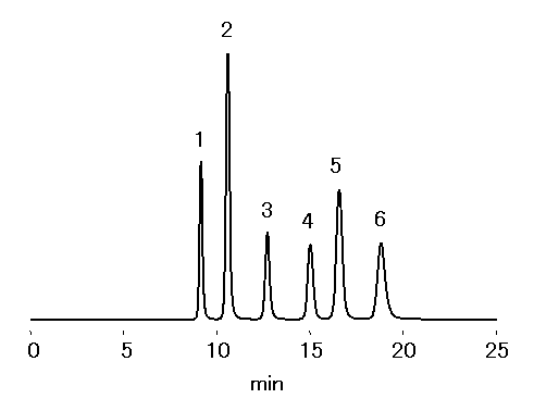
Sample : 50micro-g/mL each, 20micro-L
1. Cytosine
2. Adenine
3. Guanine
4. Uracil
5. Hypoxanthine
6. Thymine
Column       : Shodex Asahipak GS-320 HQ (7.5mmID*300mm)
Eluent       : 50mM NaH2PO4 aq./50mM H3PO4 aq.=500/380
Flow rate    : 1.0mL/min
Detector     : UV(260nm)
Column temp. : 30deg-C
Six components were completely separated using RSpak DE-413 (a column for reversed phase chromatography).
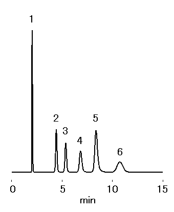
Sample : 0.01% each, 5micro-L
1. Cytosine
2. Uracil
3. Hypoxanthine
4. Guanine
5. Thymine
6. Adenine
Column       : Shodex RSpak DE-413 (4.6mmID*150mm)
Eluent       : 0.05M KH2PO4 aq.
Flow rate    : 1.0mL/min
Detector     : UV(260nm)
Column temp. : 25deg-C
Guanine and tryptophan were analyzed using three columns for reversed phase chromtography, RSpak DM-614, NN-814 and DE-613.
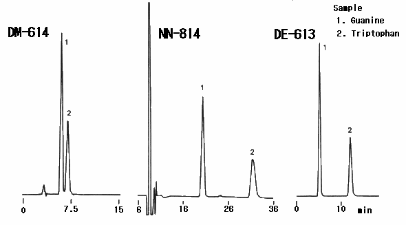
Sample : 1. Guanine , 2. Tryptophan
Columns      : (left);   Shodex RSpak DM-G (4.6mmID*10mm) + DM-614 (6.0mmID*150mm)
               (Center); Shodex RSpak NN-G (6.0mmID*50mm) + NN-814 (8.0mmID*250mm)
               (Right);  Shodex RSpak DE-G (4.6mmID*10mm) + DE-613 (6.0mmID*150mm)
Eluent       : (Left);   0.3M K2HPO4(pH8.6)
               (Center); 0.1M NaH2PO4(pH3.0 adjusted by H3PO4)
               (Right);  0.23M K2HPO4 aq./CH3OH=80/20
Flow rate    : (Left); 0.8mL/min, (Center); 1.0mL/min, (Right); 0.8mL/min
Detector     : (Left); UV(278nm), (Center); Shodex RI, (Right); UV(278nm)
Column temp. : (Left); 30deg-C, (Center); 50deg-C, (Right); 30deg-C
Adenosine and derivatives were analyzed using Asahipak GS-320 HQ (a multimode column).
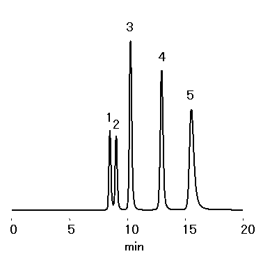
Sample : 100micro-g/mL each, 20micro-L
1. ATP
2. ADP
3. AMP
4. Adenine
5. Adenosine
Column       : Shodex Asahipak GS-320 HQ (7.5mmID*300mm)
Eluent       : 200mM NaH2PO4 aq./ 200mM H3PO4 aq.=500/62
Flow rate    : 1.0mL/min
Detector     : UV(260nm)
Column temp. : 30deg-C
Nucleosides were analyzed using Asahipak GS-320 HQ ( a multimode column ).
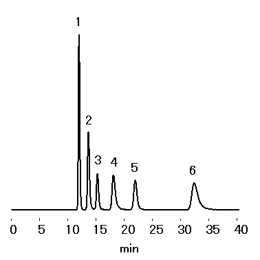
Sample : 50micro-g/mL each, 20micro-L
1. Cytidine
2. Uridine
3. Inosine
4. Thymidine
5. Guanosine
6. Adenosine
Column       : Shodex Asahipak GS-320 HQ (7.5mmID*300mm)
Eluent       : 10mM NaH2PO4 aq.
Flow rate    : 1.0mL/min
Detector     : UV(260nm)
Column temp. : 30deg-C
The ligand of AFpak APB-894 (an affinity column) is aminophenyl boronic acid which generates cyclic esther reversible with diol-type hydroxsyl group. The column is suited to highly selective analysis and fractionation of nucleic acid components.
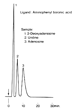
Sample :
1. 2-Deoxyadenosine
2. Uridine
3. Adenosine
Column       : Shodex AFpak APB-894 (8.0mmID*50mm)
Eluent       : 0.1M Sodium phosphate buffer(pH7.5)
Flow rate    : 1.0mL/min
Detector     : Shodex RI
Column temp. : 25deg-C
The ligand of AFpak AED-894 (an affinity column) column is ethylenediamine diacetic acid whose capacity to generate chelate is weaker than that of iminodiacetic acid. The column is suited to highly selective analysis and fractionation of nucleic acid components.
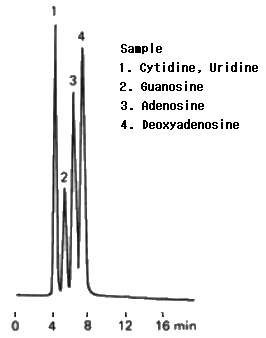
Sample :
1. Cytidine, Uridine
2. Guanosine
3. Adenosine
4. Deoxyadenosine
Column       : Shodex AFpak AED-894 (8.0mmID*50mm)
Eluent       : 0.05M 4-Ethylmorpholine-acetate buffer(pH5.65)
Flow rate    : 1.0mL/min
Detector     : UV(280nm)
Column temp. : 25deg-C
The ligand of AFpak AIA-894 (an affinity column) is iminodiacetic acid. A custom column which is co-ordinated with cupper ion was used for the analysis of ribonucleosides. First, ribonuclosides were adsorbed, and then, released by 0.05M ethylmorpholine acetate(pH6.0). The sample was separated into four peaks.
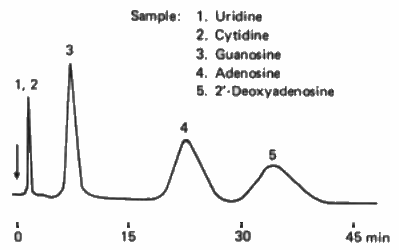
Sample :
1. Uridine
2. Cytidine
3. Guanosine
4. Adenosine
5. 2′-Deoxyadenosine
Column       : Shodex AFpak AIA-894 (8.0mmID*50mm) (Cu co-ordinated using 0.05M cupric sulfate aq.)
Eluent       : 0.05M Ethylmorpholine acetate buffer(pH6.0)
Flow rate    : 1.0mL/min
Detector     : UV(280nm)
Column temp. : Room temp.
AFpak APB-894 (an affinity column) which has aminophenyl boronic acid as the ligand was used to separate 2′-deoxyadenosine, uridine and adenosine.
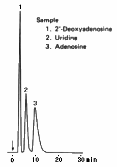
Sample :
1. 2′-Deoxyadenosine
2. Uridine
3. Adenosine
Column       : Shodex AFpak APB-894 (8.0mmID*50mm)
Eluent       : 0.1M Sodium phosphate buffer(pH7.5)
Flow rate    : 0.5mL/min
Detector     : Shodex RI
Column temp. : 25deg-C
With Asahipak GS-320 HQ (a multimode column), proteins and nucleic acid components of high molecular weights are separated by the GFC mode, they elute all together at the Vo position. One of the advantages of GS-320 HQ is, for samples consisting of low molecular weight compounds coexisting with those of high molecular weight, that the low molecular weight compounds can be analyzed directly without removing high molecular weight compounds such as proteins.
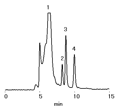
Sample : 20micro-L
1. Hemoglobin 0.85%
2. ATP 10μg/mL
3. ADP 10μg/mL
4. AMP 10μg/mL
Column       : Shodex Asahipak GS-320 HQ (7.5mmID*300mm)
Eluent       : 50mM NaH2PO4 + 50mM Na2HPO4 + 300mM NaCl
Flow rate    : 1.0mL/min
Detector     : UV(260nm)
Column temp. : 30deg-C
In the analysis of purine base and pyrimidine base with the conventional typed LC systems having injector, detector and tubes designed for the down sized analysis, conventional typed column GS-320 HQ was compared with its semi micro typed columns. Even if semi-micro columns, GS-320A series, are used for the analysis, the performance of the separation is not decreased. High-sensitivity analysis is carried out with your LC system and parts for down sized chromatography.
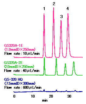
Sample : 5ppm each, 100nL
1. Cytosine
2. Uracil
3. Thymine
4. Adenine
Columns  : Shodex GS320A-1E (1.0mmID*250mm)
           Shodex GS320A-2E (2.0mmID*250mm)
           Shodex Asahipak GS-320 HQ (7.5mmID*300mm)
Eluent   : 10mM HCOONH4 buffer(pH4.0)
Detector : UV(260nm) cell volume; 3micro-L, optical path length; 7mm
Tubing   : 0.1mmID PEEK tube
Injector : Rheodyne 8125
In the analysis of uracil with the conventional typed LC systems having injector, detector and tubes designed for the down sized analysis, conventional typed column GS-320 HQ was compared with its semi-micro typed columns. The peak height of uracil analyzed with semi-micro GS320A-2E (2.0mmID) column is twelve times than that analyzed with conventional typed GS-320 HQ (7.5mmID). And the peak height of uracil analyzed with semi-micro GS320A-1E (1.0mmID) column is thirty six times than that analyzed with conventional typed GS-320 HQ (7.5mmID). The high sensitivity can be obtained with your LC system and parts designed for down sized chromatography.
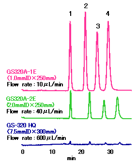
Sample : 10ppm each, 1micro-L
Uracil
Columns  : Shodex GS320A-1E (1.0mmID*250mm)
           Shodex GS320A-2E (2.0mmID*250mm)
           Shodex Asahipak GS-320 HQ (7.5mmID*300mm)
Eluent   : 10mM HCOONH4 buffer(pH4.0)
Detector : UV(260nm) cell volume; 3micro-L, optical path length; 7mm
Tubing   : 0.1mmID PEEK tube
Injector : Rheodyne 8125
The figure shows the effect of the injection volume to the separation for the analysis of uracil using GS-320 series columns.
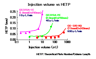
(Recommended injection volume)
| Product name |
Size (mm) ID x L |
Injection volume (micro-L) |
|---|---|---|
| GS320A-1E |
1.0 x 250 |
1
|
| GS320A-2E |
2.0 x 250
|
10
|
| GS-320 HQ |
7.5 x 300
|
100
|
Sample : Uracil
2ng (GS320A-1E)
10ng (GS320A-2E)
200ng (GS-320 HQ)
Columns  : Shodex GS320A-1E (1.0mmID*250mm)
           Shodex GS320A-2E (2.0mmID*250mm)
           Shodex Asahipak GS-320 HQ (7.5mmID*300mm)
Eluent   : 10mM HCOONH4 buffer(pH4.0)
Detector : UV(260nm) cell volume; 3micro-L, optical path length; 7mm
Tubing   : 0.1mmID PEEK tube
Injector : Rheodyne 8125
The figure shows the effect of the flow rate to the separation for the analysis of uracil using GS-320 series columns.
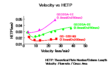
(Recommended flow rate)
|
Product name
|
Size(mm)
ID x L |
Flow rate
|
|---|---|---|
| GS320A-1E |
1.0 x 250 |
3 to 10micro-L/min
|
| GS320A-2E |
2.0 x 250
|
30 to 50micro-L/min
|
| GS-320 HQ |
7.5 x 300
|
0.4 to 1.0mL/min
|
Sample : 10ppm Uracil
0.2micro-L (GS320A-1E)
1.0micro-L (GS320A-2E)
20micro-L (GS-320 HQ)
Columns  : Shodex GS320A-1E (1.0mmID*250mm)
           Shodex GS320A-2E (2.0mmID*250mm)
           Shodex Asahipak GS-320 HQ (7.5mmID*300mm)
Eluent   : 10mM HCOONH4 buffer(pH4.0)
Detector : UV(260nm) cell volume; 3micro-L, optical path length; 7mm
Tubing   : 0.1mmID PEEK tube
Injector : Rheodyne 8125
Purine base and its final metabolite, uric acid, were separated using multi-mode column, Asahipak GS-320 HQ. Purine base is a general name for compounds (nucleotide, nucleoside, base) containing purine structure and found in most foods. Through metabolic pathways, purines are broken down to uric acid, when exceeded amount of uric acid is accumulated in body, it may cause gout.
For the analysis of purines in food, first, homogenized food is freeze-dried and then dissolved in 70%H2O2. By this hydrolysis process neutralizes the purines into purine bases.
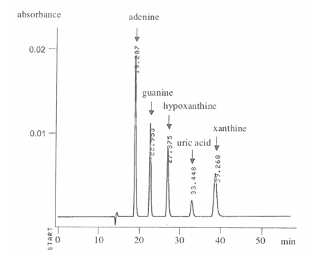
Sample : Standard purin bases
Adenine
Guanine
Hypoxanthine
Uric acid
Xanthine
Column       : Shodex Asahipak GS-320 HQ (7.5mmID*300mm)
Eluent       : 150mM Sodium phosphate buffer(pH2.5)
Flow rate   : 0.6mL/min
Detector     : UV(260nm)
Column temp. : 35deg-C
Data provided by Prof. Kiyoko Kaneko, Faculty of Pharmaceutical Sciences, Teikyo University
Purine base in beer was analyzed using multi-mode column, Asahipak GS-320 HQ. (A) Regular beer, (B) Beer processed with Guanase (guanine deaminase: enzyme converts guanine to xanthine). The result shows decrease of guanine and increase of xanthine. Purine base is a general name for compounds (nucleotide, nucleoside, base) containing purine structure and found in most foods. Through metabolic pathways, purines are broken down to uric acid, when exceeded amount of uric acid is accumulated in body, it may cause gout. For the analysis of purines in food, first, homogenized food is freeze-dried and then dissolved in 70%H2O2. By this hydrolysis process neutralizes the purines into purine bases.
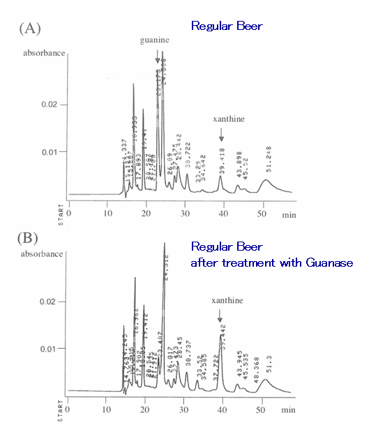
Column       : Shodex Asahipak GS-320 HQ (7.5mmID*300mm)
Eluent       : 150mM Sodium phosphate buffer(pH2.5)
Flow rate   : 0.6mL/min
Detector     : UV(260nm)
Column temp. : 35deg-C
Data provided by Prof. Kiyoko Kaneko, Faculty of Pharmaceutical Sciences, Teikyo University

